




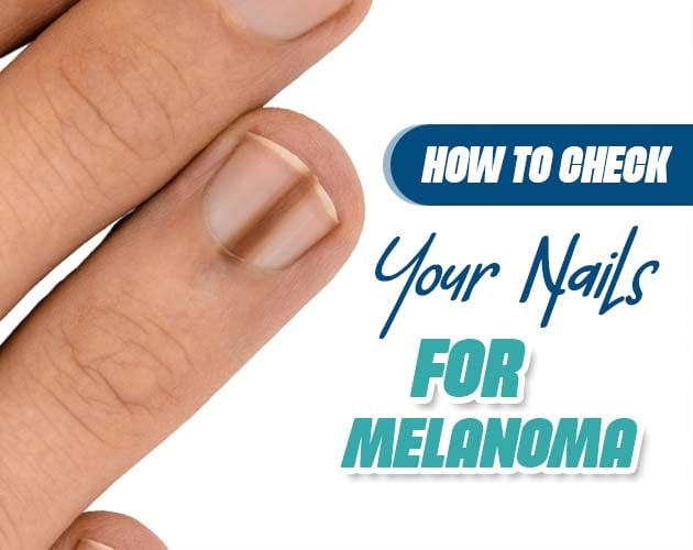
When checking your skin for suspicious moles, nails are frequently overlooked. However, skin cancer can develop beneath fingernails and toenails in a rare but serious form known as subungual melanoma.
Unlike cutaneous melanomas triggered primarily by ultraviolet (UV) radiation from sunlight or tanning beds, subungual malignancy originates in the nail matrix, the tissue under the nail bed where keratinized nail cells are produced. Because it often mimics common nail conditions like fungal infections or bruises from trauma, early detection requires knowing precisely what visual signs to look for.
Understanding Subungual Melanoma and Its Risk Factors
Subungual melanoma accounts for roughly 0.7% to 3.5% of all melanoma cases worldwide. While relatively rare in light-skinned populations, it represents a significant proportion of melanoma diagnoses in individuals with darker skin tones, including people of African, Asian, and Hispanic descent.
Unlike typical skin malignancies, subungual cases are not strongly associated with sun exposure. Instead, potential risk factors include:
- Prior Physical Trauma: Repeated micro-trauma or significant acute injury to the nail unit (though trauma can also simply draw attention to an existing lesion).
- Personal or Family History: Individuals with a history of dysplastic nevi or cutaneous malignancies.
- Age: Diagnoses occur most frequently between the ages of 50 and 70.
Pathological Mechanism:
[Atypical Melanocyte Proliferation in Nail Matrix] ➔ [Pigment Transfer into Keratinized Cells] ➔ [Pigmented Band Grows Outward with Nail Plate]
Visual Indicators: The ABCDEF Rule for Nail Health
Dermatologists use a modified mnemonic framework to help patients and clinicians evaluate pigmented nail bands (longitudinal melanonychia).
The ABCDEF Diagnostic Framework:
- A – Age & Ethnicity: Highest incidence in individuals aged 50–70 and populations with darker skin phototypes.
- B – Band Characteristics: A pigmented streak that is Brown or Black, wide (3 mm), or features irregular, varying color tones.
- C – Change: Rapid expansion, darkening, or changes in the shape or growth rate of the streak over time.
- D – Digit Involved: Most commonly affects the primary digits the thumb, great toe (index digit), or index finger.
- E – Extension (Hutchinson’s Sign): Pigment spreading beyond the nail plate onto the surrounding cuticle or proximal nail fold (hyponychium). This is a strong clinical indicator of subungual malignancy.
- F – Family or Personal History: A prior history of atypical moles or melanoma.
Differential Diagnosis: Subungual Melanoma vs. Common Nail Conditions
Many dark lines on nails are completely benign. Recognizing how subungual malignancies differ from everyday nail issues prevents unnecessary anxiety while ensuring timely medical evaluation.
| Feature | Subungual Melanoma | Subungual Hematoma (Bruise) | Fungal Infection (Onychomycosis) | Benign Longitudinal Melanonychia |
| Primary Cause | Atypical melanocyte growth | Physical injury / Traumatic bleeding | Fungal pathogen | Benign nevus or ethnic pigmentation |
| Band Color | Dark brown to black with uneven shades | Red, purple, or dark brown/black | Yellow, white, or greenish brown | Uniform light brown to dark brown |
| Movement with Growth | Persists at matrix as nail grows outward | Moves outward with nail growth over time | Spreads irregularly, causing nail crumbling | Stationary, remains stable for years |
| Cuticle Pigment | Often present (Hutchinson’s sign) | Absent | Absent | Absent |
| Nail Destruction | May cause splitting or dystrophy | Rare (unless severe acute trauma) | Common (thickening and lifting) | Absent |
How to Conduct a Systematic Home Nail Check?
Conducting a monthly self-inspection takes only a few minutes and allows you to catch subtle changes early.
Step 1: Remove All Nail Polish
Thick lacquers, gels, and acrylics completely obscure the nail matrix and bed. Always inspect bare fingernails and toenails between manicures or pedicures.
Step 2: Examine Under Bright Lighting
Inspect each digit under direct light, paying close attention to:
- The thumb and great toe.
- The proximal nail fold (cuticle area).
- The undersides of the free edge of the nail.
Step 3: Check for Longitudinal Bands
Look for vertical dark lines running from the cuticle up to the tip. Note the width, color consistency, and whether multiple nails display similar streaks (multiple streaked nails are more commonly ethnic melanonychia rather than a solitary tumor).
Step 4: Look for Cuticle Discoloration
Check if pigment bleeds past the edge of the nail matrix onto adjacent skin. For a detailed step-by-step breakdown on conducting home assessments and evaluating streak characteristics, you can read the guide on how to check your nails for melanoma by Sayyal Health.
When to Consult a Dermatologist
You should schedule a professional examination if you observe any of the following clinical signs:
- A single dark streak appears suddenly on a fingernail or toenail without prior injury.
- An existing streak grows wider than 3 millimeters or develops varying colors.
- The nail plate splits, lifts from the nail bed (onycholysis), or becomes dystrophic without an obvious cause.
- Pigment extends onto the surrounding cuticle or skin (Hutchinson’s sign).
- A bruise under the nail does not move outward as the nail grows over 2 to 3 months.
During a consultation, a clinician will use a dermatoscope, a specialized magnifying instrument to examine the pigment patterns in detail. If suspicion remains, a small biopsy of the nail matrix is performed to analyze tissue at the microscopic level.
Frequently Asked Questions
Can a subungual hematoma (bruise) turn into melanoma?
No. A subungual hematoma is a collection of blood caused by broken blood vessels following trauma, whereas melanoma is a genetic alteration in pigment-producing cells. However, people often misinterpret an early tumor as an unremembered bruise, which is why monitoring whether the discoloration grows out with the nail is essential.
Is subungual melanoma caused by UV light from gel manicure lamps?
While nail lamps emit UVA rays, subungual cases develop deep within the matrix shielded by the nail plate and are not typically linked to UV exposure. However, using broad-spectrum sunscreen or UV-blocking gloves during gel manicures remains a good skin safety practice for protecting the dorsal hands.
What is amelanotic subungual melanoma?
Amelanotic lesions lack melanin pigment entirely. Instead of appearing as a brown or black streak, they present as a red, flesh-colored, or ulcerated nodule under the nail, often mimicking a non-healing wound, pyogenic granuloma, or ingrown nail.
How fast does subungual melanoma progress?
In situ forms (confined to the epidermis of the matrix) can remain stable or progress slowly over months to years. Invasive forms can progress more rapidly, which highlights why early biopsy of suspicious longitudinal streaks is critical for a favorable prognosis.
Summary
Subungual melanoma is a rare but manageable condition when identified in its early stages. By performing regular bare-nail self-checks, using the ABCDEF criteria, and consulting a dermatologist whenever a dark streak expands or causes cuticle discoloration, you can protect your long-term health and ensure timely intervention.